Inside the Breathing Machine: How VReaLab Brings the Respiratory System to Life Through Immersive VR
The respiratory system is a masterpiece of biological engineering, yet students have long relied on flat textbook diagrams to understand its three-dimensional elegance. VReaLab Biology Body Structure delivers an unprecedented visual journey into respiratory anatomy, turning biology interactive learning into hands-on exploration.
Every minute of every day, an intricate mechanical and chemical performance is taking place inside each of us. Without conscious effort, the respiratory system orchestrates the movement of air through a series of precisely shaped passageways, exchanges oxygen and carbon dioxide at the cellular level, and sustains the metabolic processes that keep every tissue in the body alive. For generations of biology students, however, this remarkable system has been presented primarily through flat textbook diagrams — a representation that, however well-illustrated, cannot do justice to the three-dimensional elegance of the human body. VReaLab Biology Body Structure changes this reality. As a comprehensive 3d anatomy of the human body, this platform delivers an unprecedented visual journey into the structure of the respiratory system, transforming biology interactive learning into a hands-on exploration of form, proportion, and spatial relationship.
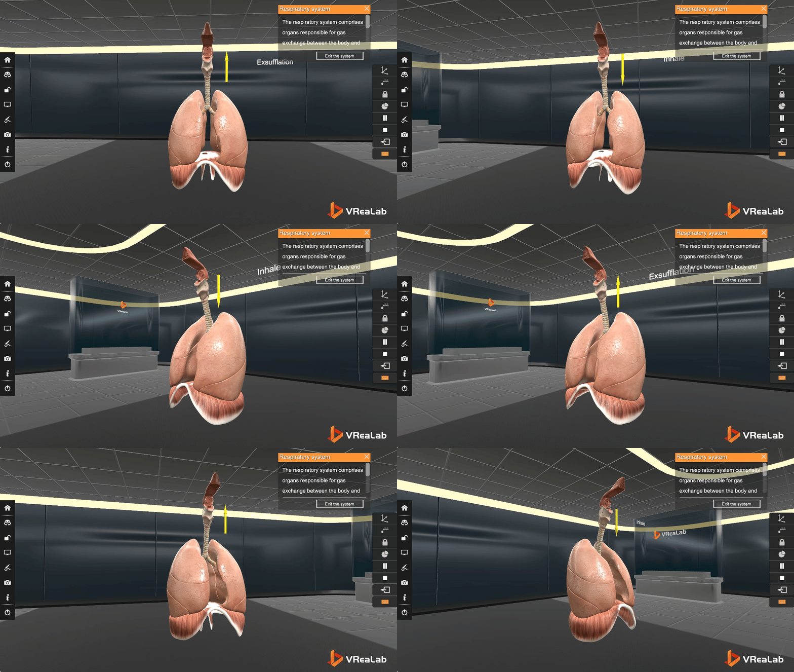
A System Built in Layers: From Airways to Alveoli
The respiratory system is a masterpiece of biological engineering — a stepwise series of increasingly smaller passages that begin with the nose and mouth, descend through the pharynx and larynx, and travel down the trachea before branching into the bronchi and bronchioles, finally terminating in the delicate alveolar sacs where gas exchange occurs. Each level of this system has a unique structure perfectly suited to its function. With VReaLab's Human Body Systems 3D Model, students can examine each of these layers with unprecedented clarity — zooming in, rotating, and isolating individual structures to understand exactly how form follows function at every scale.
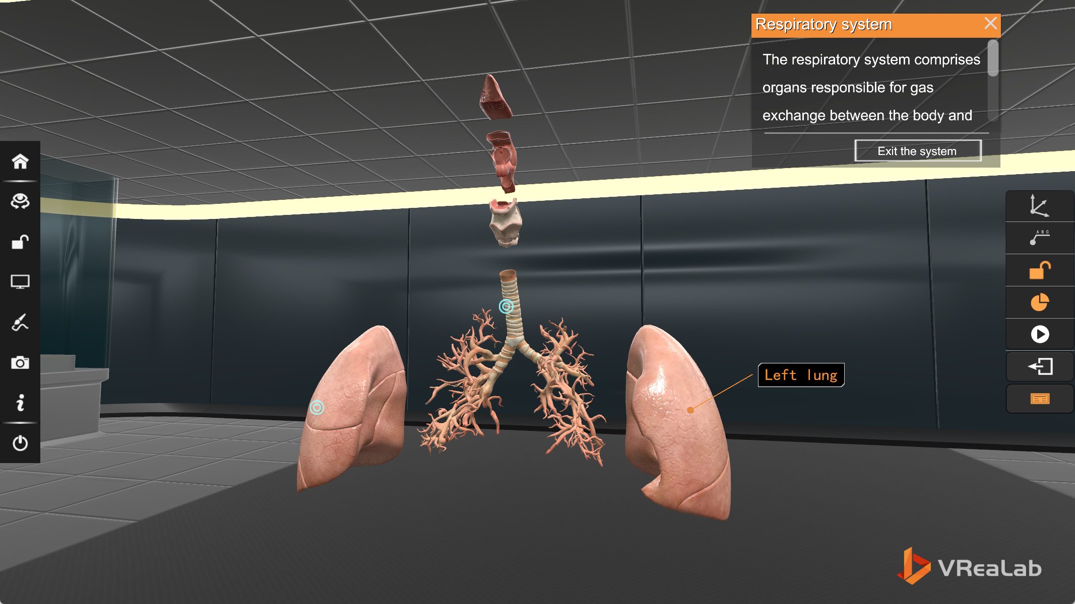
The image below shows the full respiratory system in its assembled form: the left lung displayed alongside the intricate branching of the bronchial tree, the trachea descending from the laryngeal complex above. VReaLab lets students interact with this scene freely — selecting individual structures, adjusting transparency to reveal deeper anatomical layers, and exploring the spatial relationships that no static illustration can convey.
The Lungs and Bronchial Tree: A Closer Look
Step closer into the VReaLab scene and the lungs reveal their full anatomical story. The right and left lungs are not identical — the right lung is divided into three lobes, while the left lung has only two, accommodating the heart that sits slightly to the left of the midline. This kind of detail, often glossed over in textbooks, becomes immediately apparent when students can freely rotate the model and observe the asymmetry for themselves. Below the trachea, the primary bronchi branch into secondary and tertiary bronchi, then into progressively smaller bronchioles, ending in alveolar clusters that together provide roughly 70 square meters of surface area for gas exchange.
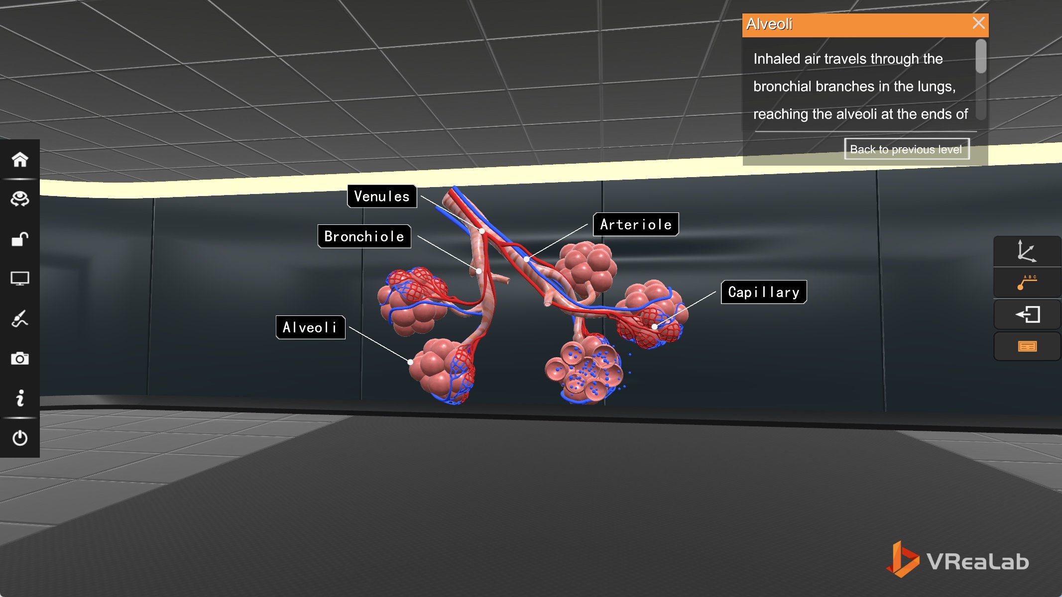
Through VReaLab's biology interactive learning environment, students can select either lung to view it from any angle. The label system — clearly shown pointing to the right lung in the image below — reveals not just the name of each structure, but its position, its boundaries, and its connection to neighboring organs. By clicking on any organ, students trigger contextual information panels that describe both the structure and its physiological role.
This is biology interactive learning at its best — a system that responds to student curiosity by allowing free exploration, selection, and manipulation of every anatomical element. Rather than reading about the right lung in isolation, students see it in context: positioned against the chest wall, anchored by the mediastinum, and connected to the trachea above. The 3d anatomy of the human body, in this sense, is not just a visual experience but a spatial and tactile one — one that builds durable mental models far more effectively than rote memorization of Latin terminology.
From Classroom to Inquiry: Reimagining How Biology Is Taught
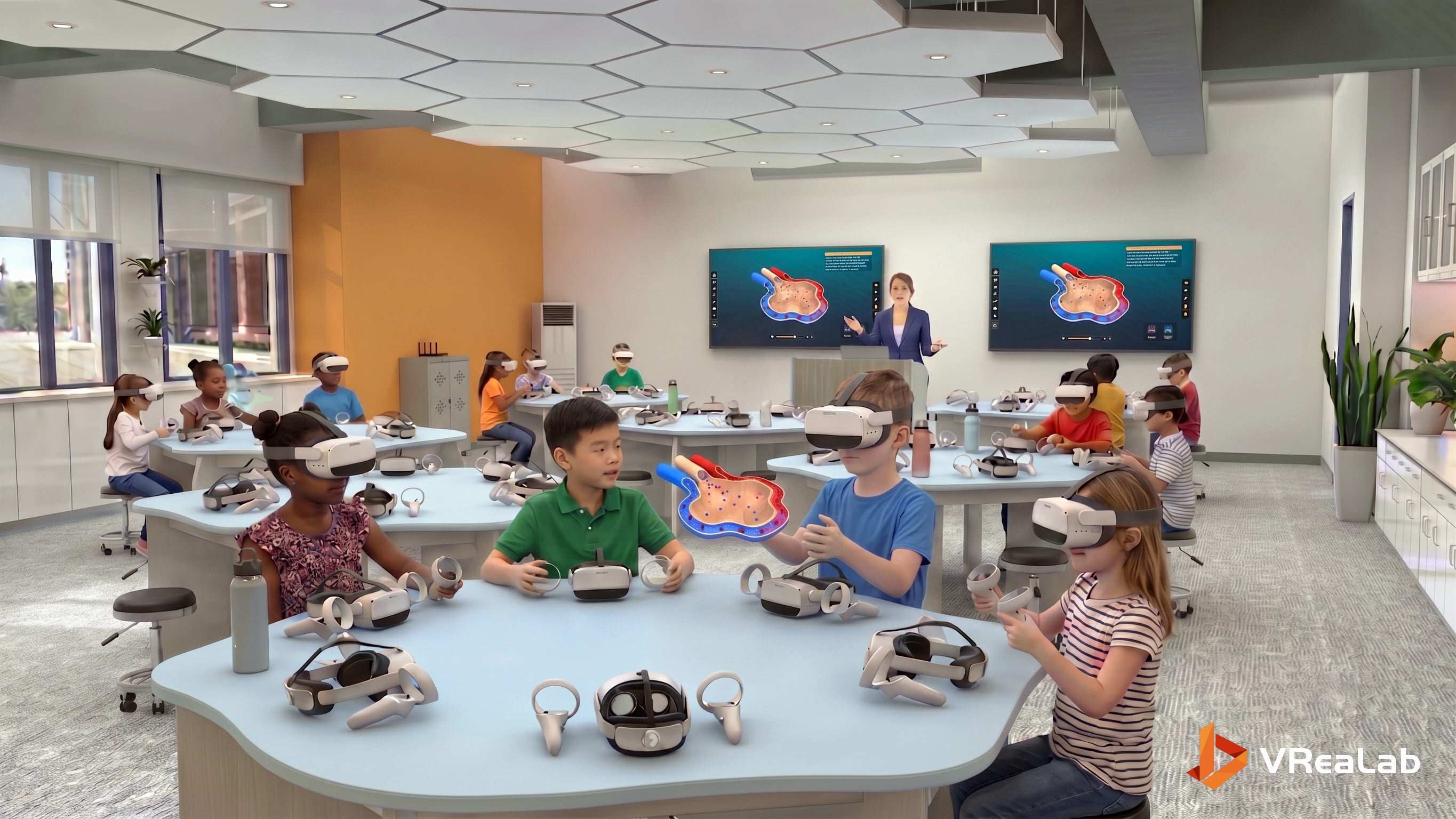
VReaLab's impact extends well beyond the screen. In classrooms equipped with VR headsets, students gather around the digital model as they once gathered around a dissection tray — but without the safety concerns, ethical limitations, or one-time-use constraints of physical specimens. The image below captures this transformation in action: students wearing VR headsets, exploring the respiratory system from multiple angles, with supplementary materials and molecular models arranged around the table to support deeper inquiry.
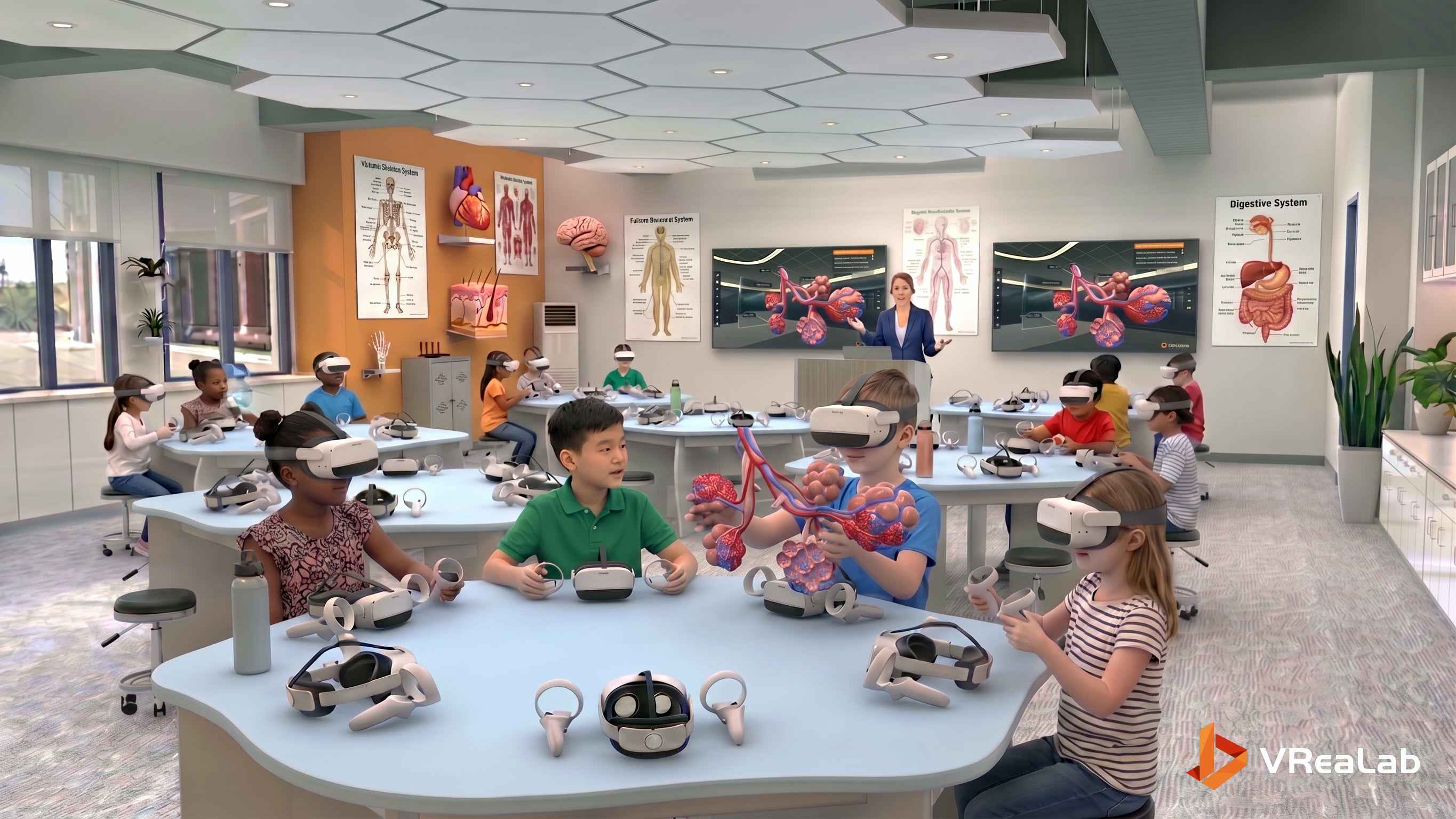
Notice the posters on the walls — visible labels such as "ALVEOLAR GAS EXCHANGE" and "STEM MECHANICS OF BREATHING" hint at the broader curriculum that VReaLab supports. The molecule models on the table (O₂, CO₂, and related structures) provide a tangible complement to the immersive VR experience, allowing students to bridge the macroscopic world of organs with the microscopic world of molecules. This multi-modal approach — combining VR headsets, physical models, and collaborative discussion — represents the future of biology interactive learning, where technology amplifies rather than replaces the hands-on, social nature of science education.
Conclusion
The respiratory system is one of the most important and elegant systems in the human body, and yet for decades, students have struggled to understand it through two-dimensional images and static diagrams. VReaLab Biology Body Structure changes that. By providing a complete, interactive, and immersive 3d anatomy of the human body, the platform transforms biology interactive learning from a passive experience into an active journey of discovery. Whether examining the branching pattern of the bronchial tree, comparing the asymmetry of the two lungs, or simply marveling at the architectural elegance of a system that sustains life with every breath, students using VReaLab's Human Body Systems 3D Model gain an understanding that is not just deeper — but more lasting, more meaningful, and far more enjoyable.