Understanding Respiratory Function Through VReaLab’s Immersive VR Visualization
Most people rarely think about breathing — it happens automatically, roughly 20,000 times per day. Yet behind every breath lies an extraordinary sequence of mechanical and biochemical events. VReaLab Biology Body Function provides an interactive, animated view of every step, transforming biology interactive learning into a vivid exploration of how the respiratory system actually works.
Most people rarely think about breathing. It happens automatically — roughly 20,000 times per day — yet behind every breath lies an extraordinary sequence of mechanical and biochemical events. The diaphragm contracts and flattens, the rib cage expands, pressure inside the thoracic cavity drops, air rushes in through the nose and trachea, and ultimately oxygen molecules cross into the bloodstream at the alveolar surface. For biology students, understanding this process has always been a challenge: it is dynamic, three-dimensional, and involves structures that are difficult to see. VReaLab Biology Body Function changes this by providing an interactive, animated view of every step. As a comprehensive 3d anatomy of the human body with a dedicated focus on physiological processes, VReaLab transforms biology interactive learning into a vivid exploration of how the respiratory system actually works.
The Mechanics of Breathing: A Living Animation
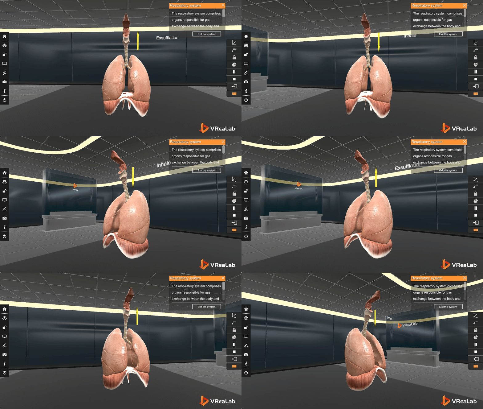
Breathing is one of the most elegant examples of biomechanics in the human body. It is driven primarily by the diaphragm — a dome-shaped muscle that separates the thoracic cavity from the abdominal cavity — along with the intercostal muscles that lie between the ribs. When the diaphragm contracts, it flattens and descends, increasing the volume of the thoracic cavity. Simultaneously, the external intercostal muscles contract, lifting the rib cage upward and outward. Together, these actions lower intrathoracic pressure and draw air into the lungs. Exhalation, in healthy resting breathing, is largely passive: the muscles relax, the elastic recoil of the lungs and chest wall pushes air back out.
VReaLab’s Human Body Systems 3D Model brings this mechanism to life through a dedicated Respiratory Movement module. The image below shows the in-software visualization: a transparent rib cage with labeled structures — Rib, Lung, Intercostal Muscle, and Diaphragm — alongside the lungs. Students can play, pause, and step through the breathing cycle frame by frame, watching the diaphragm descend and rise, observing the rib cage expand and contract. This is biology interactive learning at its most compelling: not a static diagram to be memorized, but a living, breathing model that responds to student exploration.
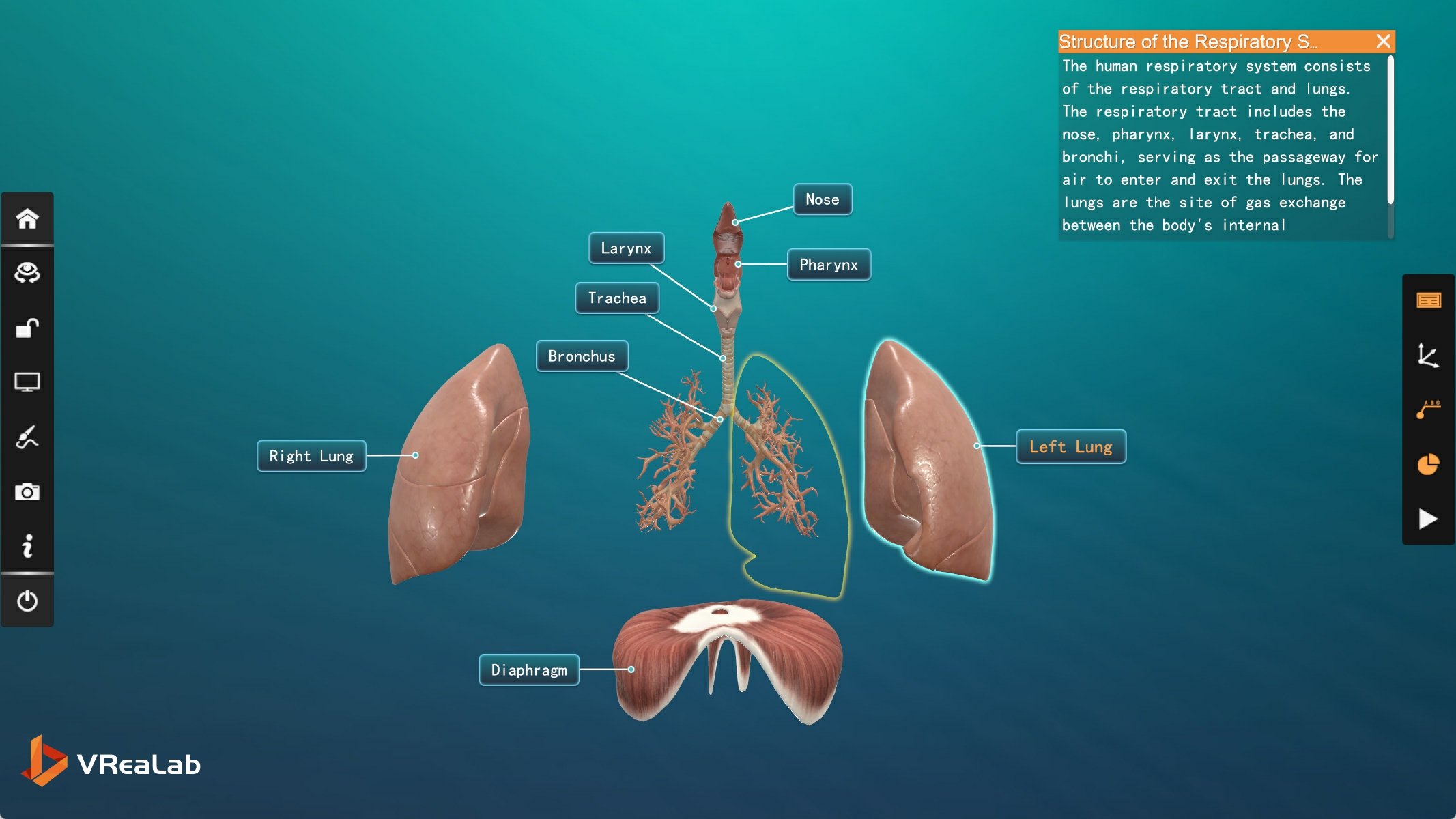
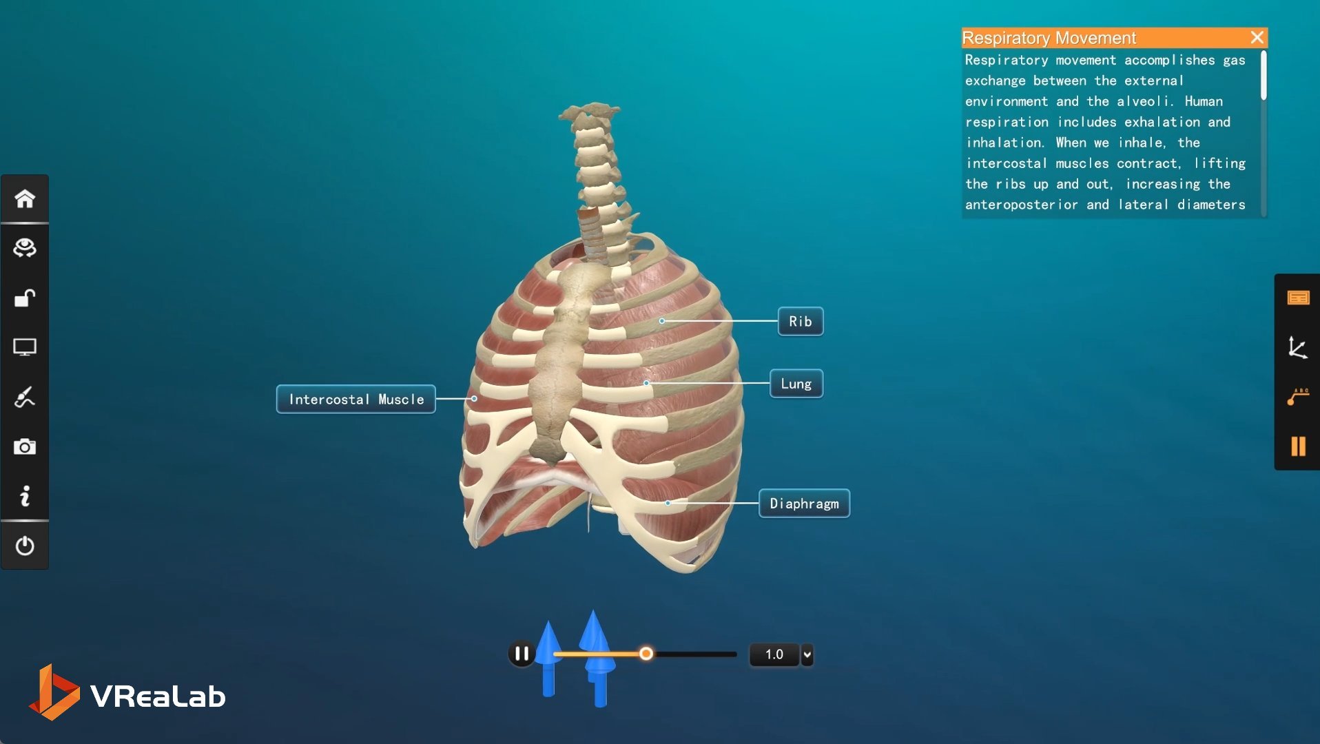
Notice the carefully placed labels in the scene. Each structure is identified and connected by a clear line to its anatomical position — the ribs forming the protective cage, the lungs filling the thoracic space, the intercostal muscles weaving between the ribs, and the diaphragm as the muscular floor below. The accompanying text panel provides a written explanation, while the playback controls below the model let students manipulate time itself: slow the breath down, speed it up, or replay specific moments. This combination of visualization, annotation, and temporal control turns an abstract physiological process into something concrete and explorable.
From Structure to Function: The Logic of the Airway
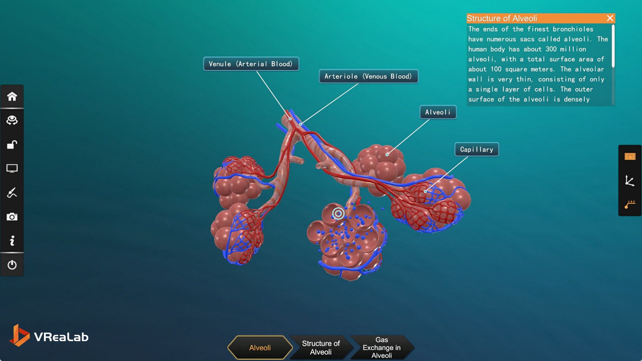
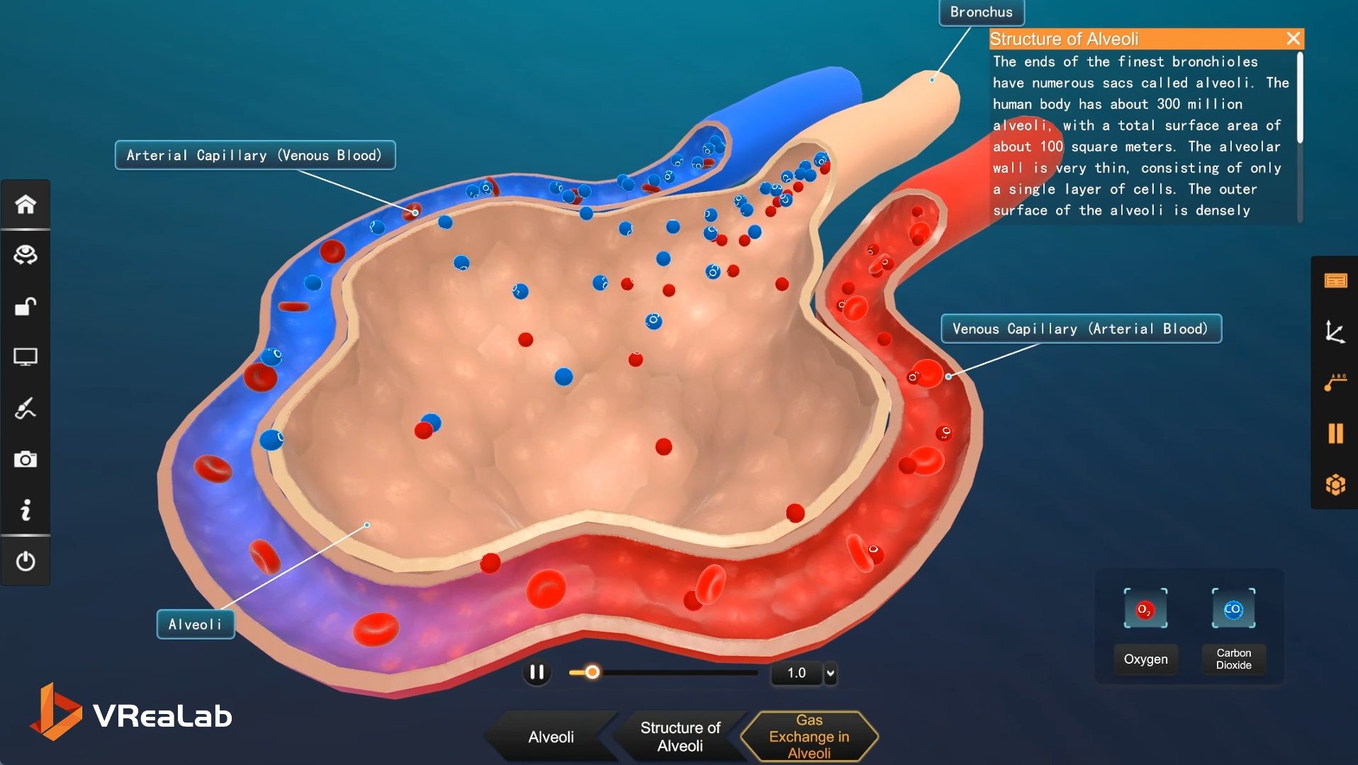
Once students have understood the mechanical basis of breathing, the next question is: what happens to the air once it gets inside? VReaLab guides learners through the entire respiratory pathway — from the nasal cavity, where air is warmed and humidified, through the larynx and trachea, into the branching bronchi, and finally to the alveolar sacs. At each stage, the anatomy is purpose-built for its function: the nasal cavity’s turbinates create turbulent airflow that warms and filters incoming air; the trachea’s C-shaped cartilage rings keep the airway permanently open; the bronchi’s tapering diameters prepare air for its ultimate destination.
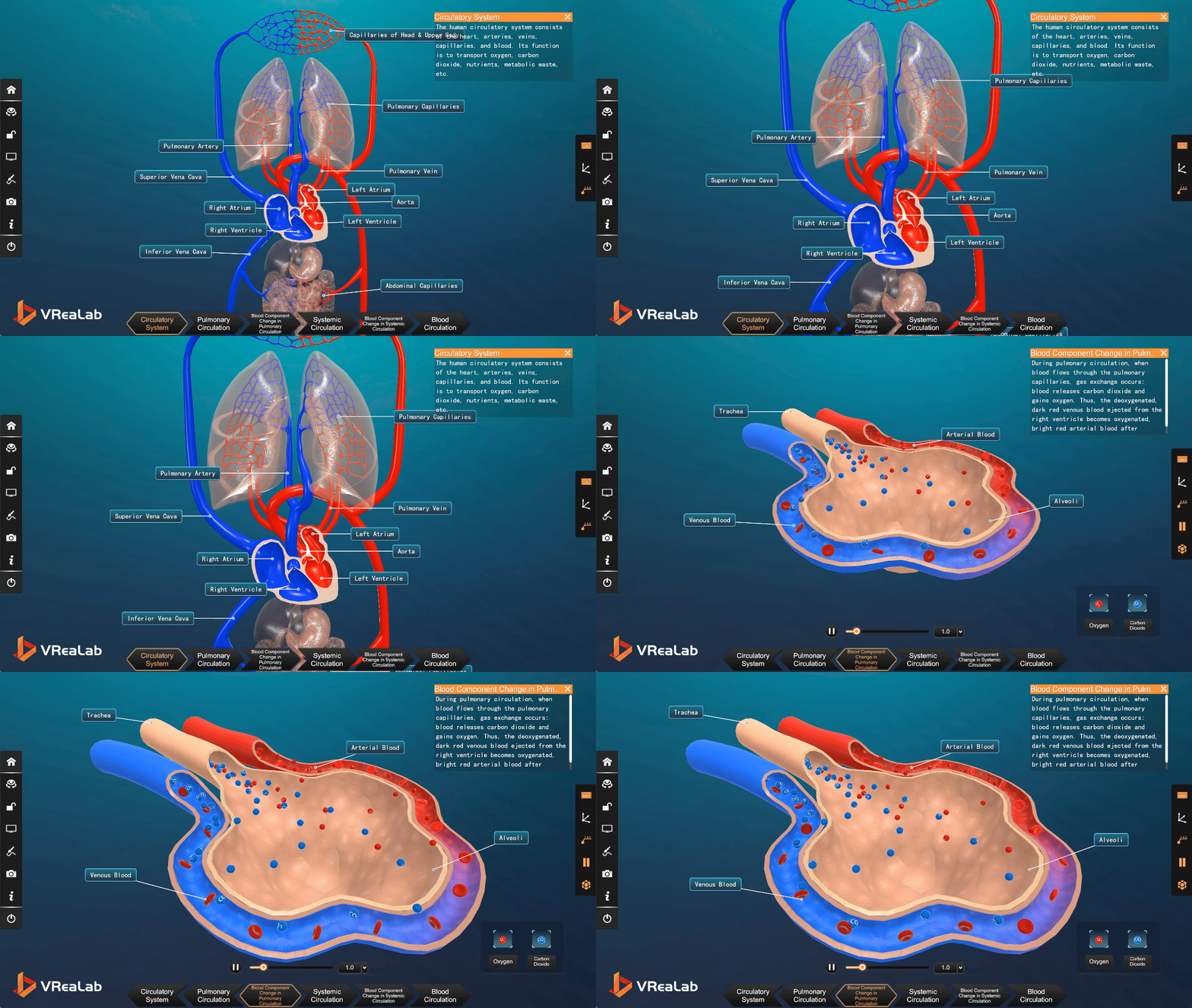
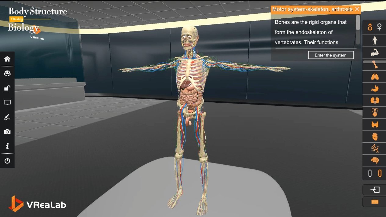
That destination is the alveolus — a tiny, balloon-like sac wrapped in an extraordinarily thin membrane and surrounded by a dense capillary network. It is here, across a barrier less than one micrometer thick, that oxygen diffuses into the blood and carbon dioxide diffuses out. This is the central exchange of life, and yet in conventional biology classrooms it is almost impossible to show. Through VReaLab’s 3d anatomy of the human body, students can trace the entire journey of a single breath — following an oxygen molecule from the moment it enters the nose to the instant it crosses the alveolar membrane and binds to a hemoglobin molecule in a red blood cell. This end-to-end view of the respiratory function is something that no textbook, model, or video has ever been able to provide.
Bringing the Function to the Classroom
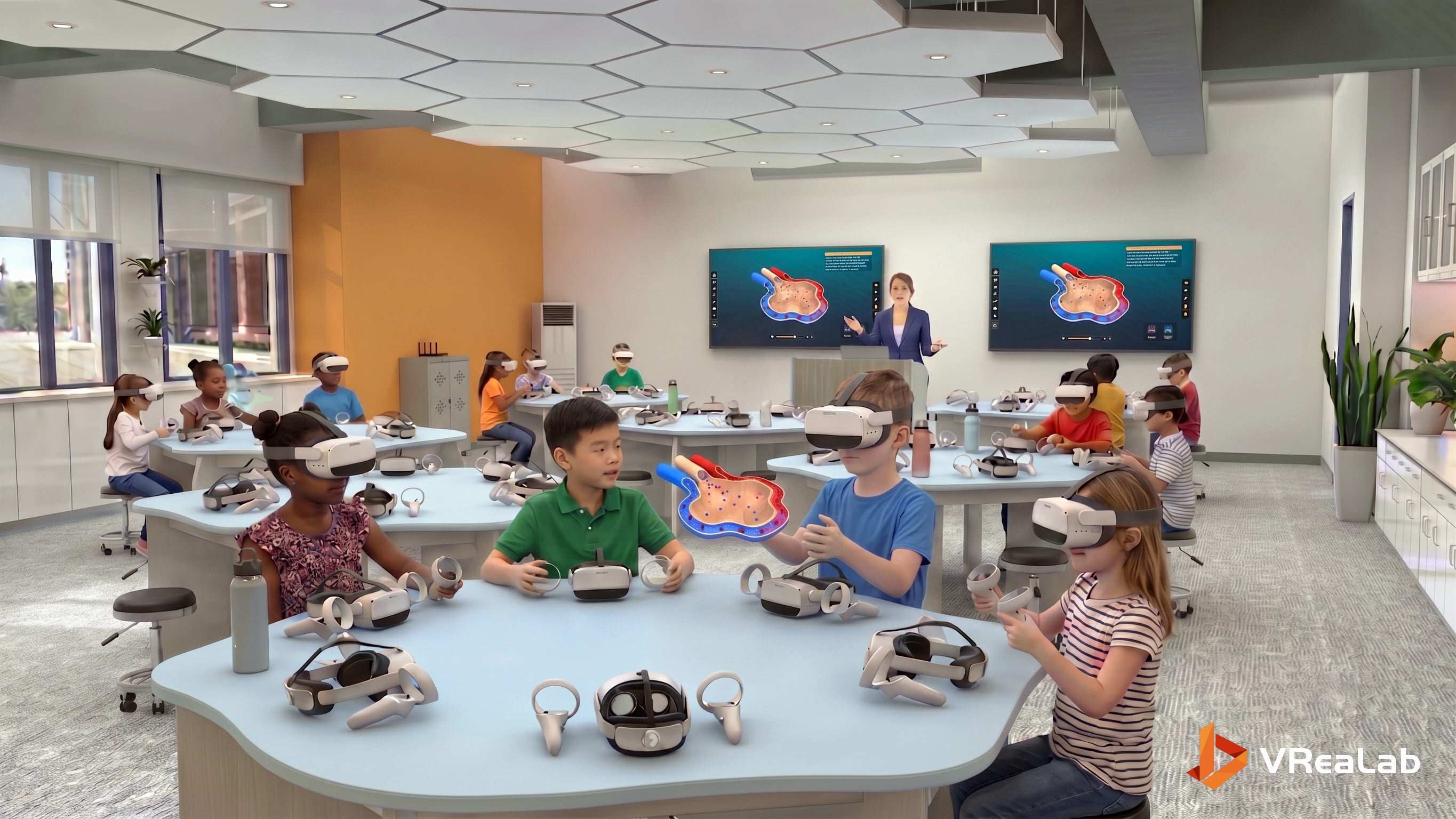
The classroom image below captures VReaLab’s impact on biology interactive learning in vivid detail. Students sit together, each wearing a VR headset, exploring the respiratory system from individual perspectives. On the central table stands a tangible model of the rib cage — a physical bridge between the virtual and the real — surrounded by molecular models (O₂, CO₂, and more) that reinforce the chemical exchange at the heart of respiration. Posters on the walls (“Alveolar Gas Exchange,” “Stem Mechanics of Breathing,” “Anatomie der Atmungswege”) signal that this is a multi-disciplinary learning environment where biology, chemistry, and even physics converge.
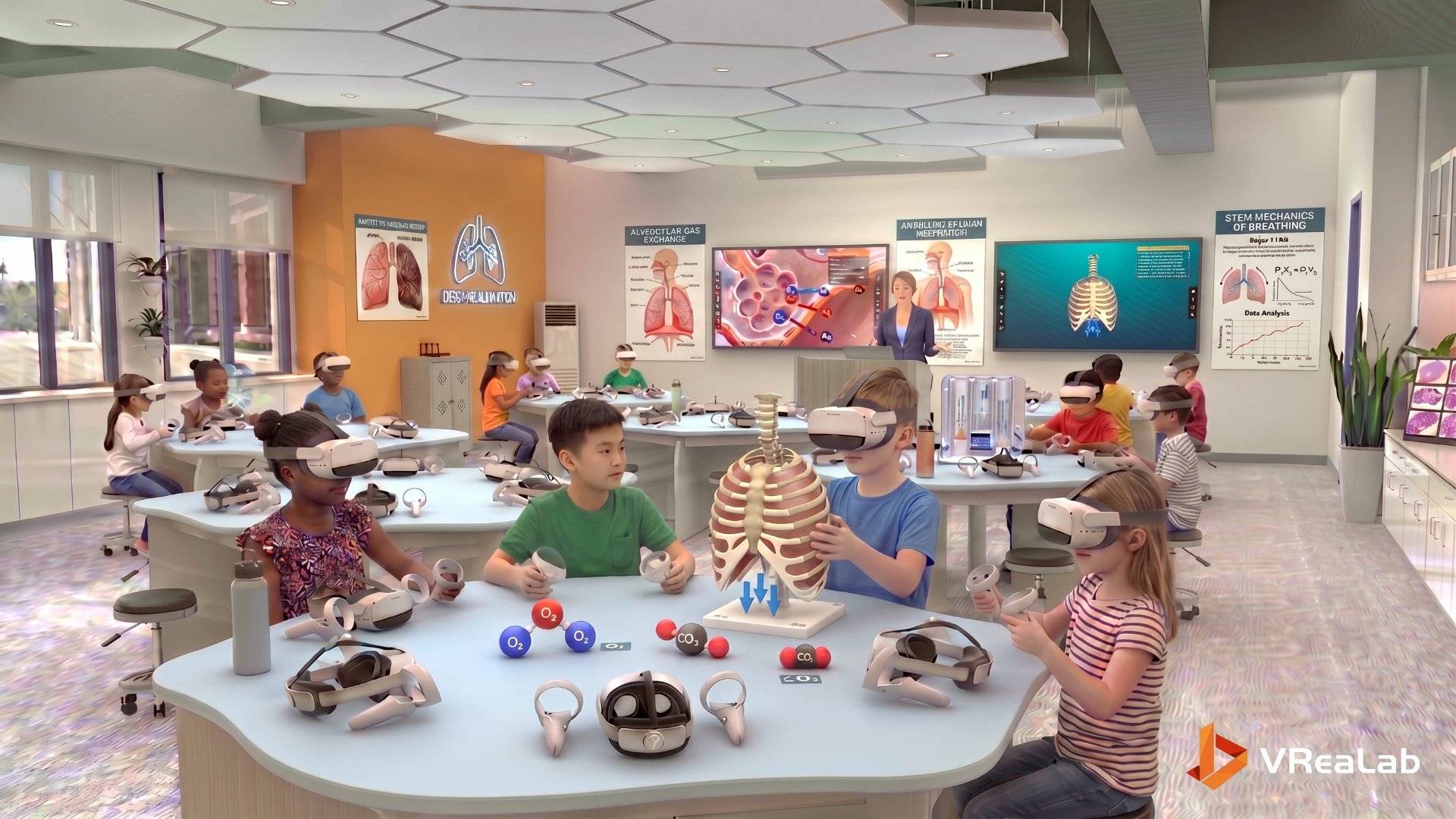
This is exactly what biology interactive learning should look like. Students are not passively watching a screen; they are actively engaged with a rich, multi-sensory environment in which the respiratory system is simultaneously explored as a 3d anatomy of the human body (structural), as a dynamic process (functional), and as a phenomenon at the molecular scale (chemical). The teacher’s role shifts from lecturing to facilitating inquiry, asking questions that guide students toward deeper understanding. Discussion becomes richer because every student has experienced the same scene from a unique angle, and can contribute a unique perspective.