Walking into the Heart: A VR Physiology Learning Experience
The heart is one of the most important organs in the human body. With virtual reality entering science education, learning about the heart is shifting from identifying structures on a page to entering the system and understanding how it works.
The heart is one of the most important organs in the human body, and also one of the easiest to misunderstand. In the textbook, students learn that it is an organ about the size of a fist, with four chambers, responsible for pumping blood through the whole body. Yet for many learners, this knowledge stays at the level of labels and diagrams: the heart keeps beating, but it is never truly understood. With virtual reality entering science education, learning about the heart is shifting from identifying structures on a page to entering the system and understanding how it works. In the biology virtual simulation laboratory, using VR software for human physiological structure and function, learners can walk into the heart from a first-person perspective and observe this precise and efficient living system directly.
From Flat Diagrams to a Three-Dimensional Living System
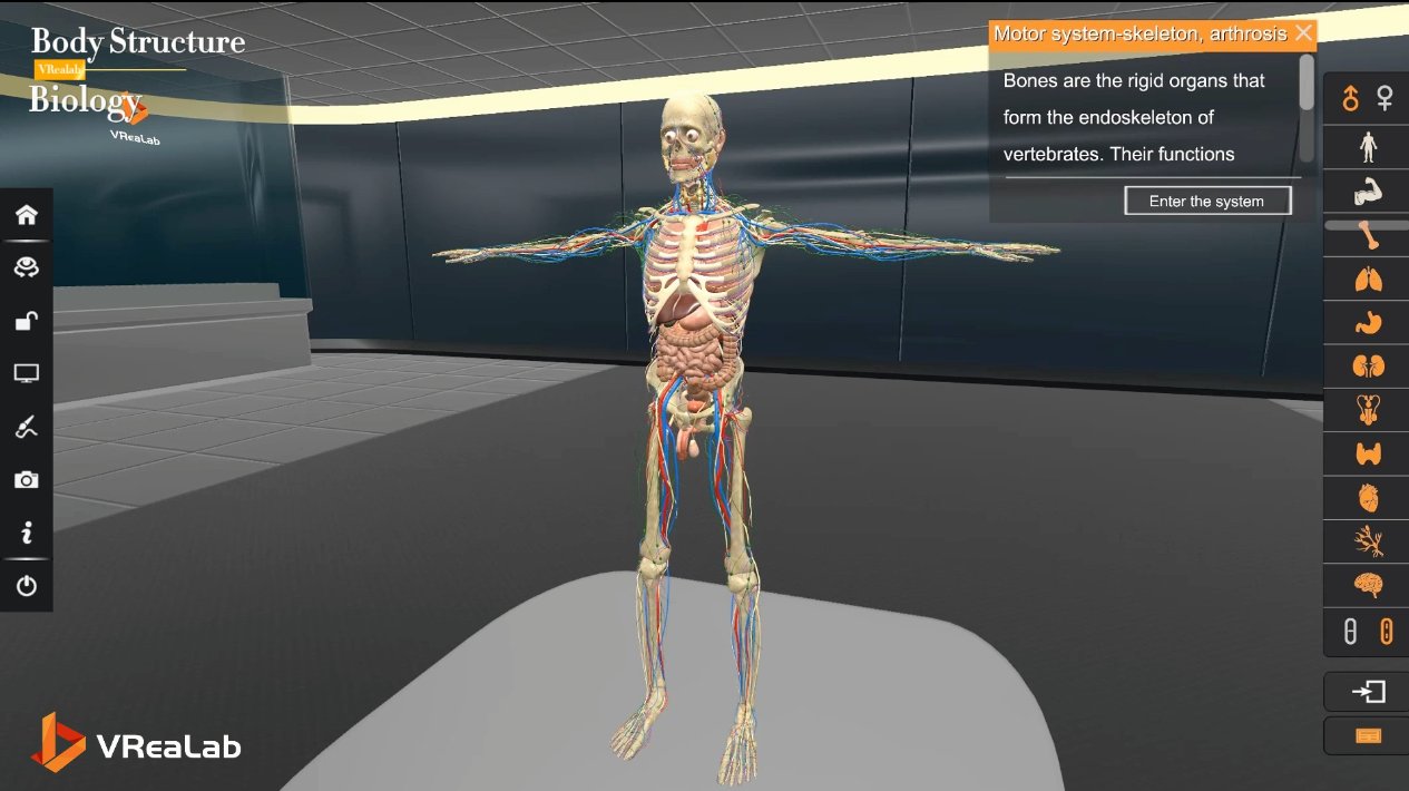
In traditional learning, the heart usually appears as a flat anatomical illustration: left atrium, right ventricle, valves, vessels. The information is complete, but it is hard for students to grasp the spatial relationships among these structures, and harder still to connect structure with function. In a VR classroom, learners enter a highly realistic 3d anatomy of the human body. The spatial position of the atria and ventricles is clear at a glance; the connection between the heart and the aorta, the pulmonary artery, and the venae cavae is presented directly; the location and direction of each valve can be observed up close. This three-dimensional, rotatable, observable way of learning helps students quickly build the cognitive framework of whole, part, and system that a printed page can never provide.
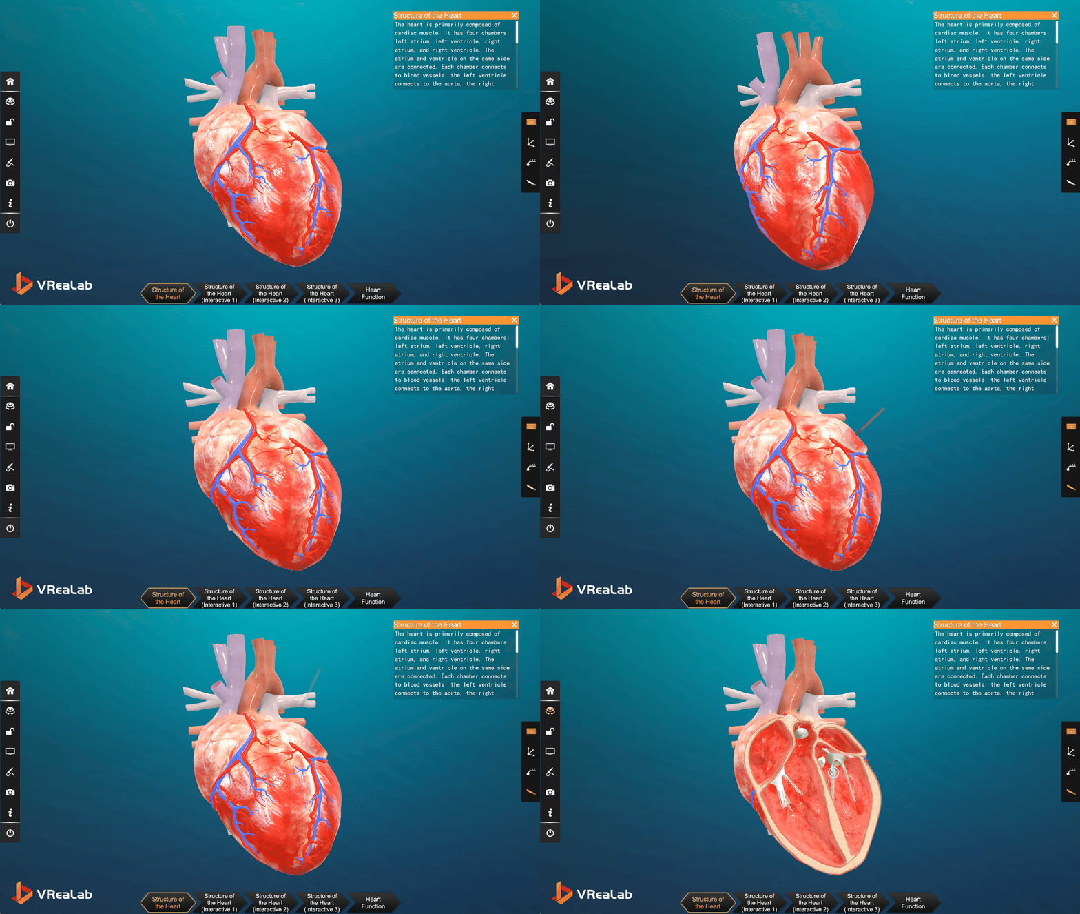
Figure 1. External structure of the heart: the aorta, pulmonary vessels, and venae cavae labeled in an interactive 3D scene.
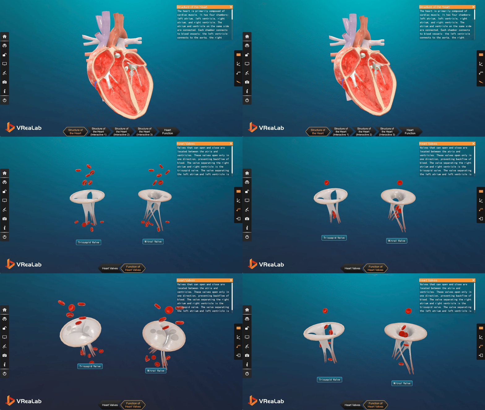
Figure 2. A sectional view of the four chambers, revealing the interior geometry that flat diagrams can only suggest.
The Heart Has Rhythm and Function, Not Just Structure
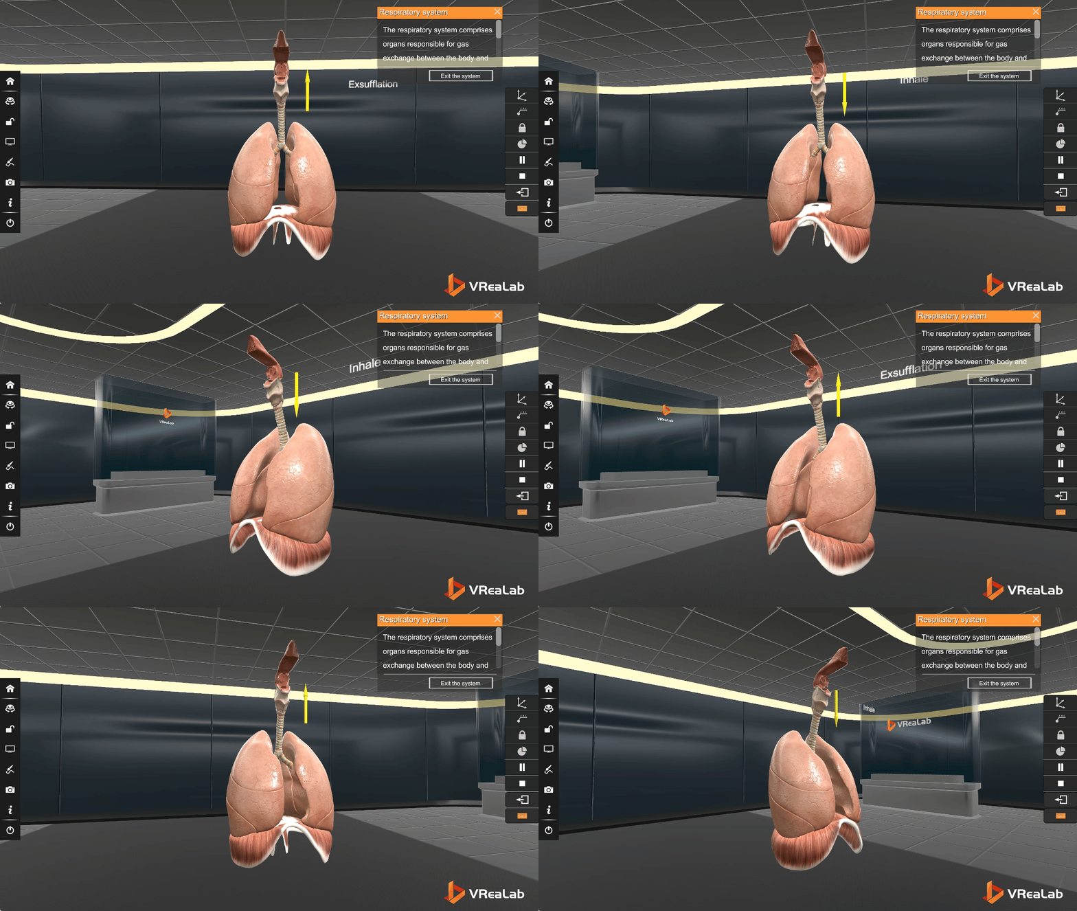
Truly understanding the heart does not come from memorizing names; it comes from seeing how it works. In the VR course, the heart is no longer a static model but a continuously running living system. Learners can observe the rhythmic sequence of atrial and ventricular contraction and relaxation, watch the direction of blood flow inside the chambers, and compare the paths of arterial and venous blood in different cavities. A close-up of the cardiac valves makes the key mechanism visible: the tricuspid and mitral valves open in only one direction, which is exactly why blood never flows backward. Through this kind of biology interactive learning, students naturally connect structure with function, and the valve stops being a vocabulary word and becomes a working piece of engineering they have seen in action. Because the whole scene can be slowed down and replayed, even the rapid sequence of a single heartbeat becomes an observable, discussable process.
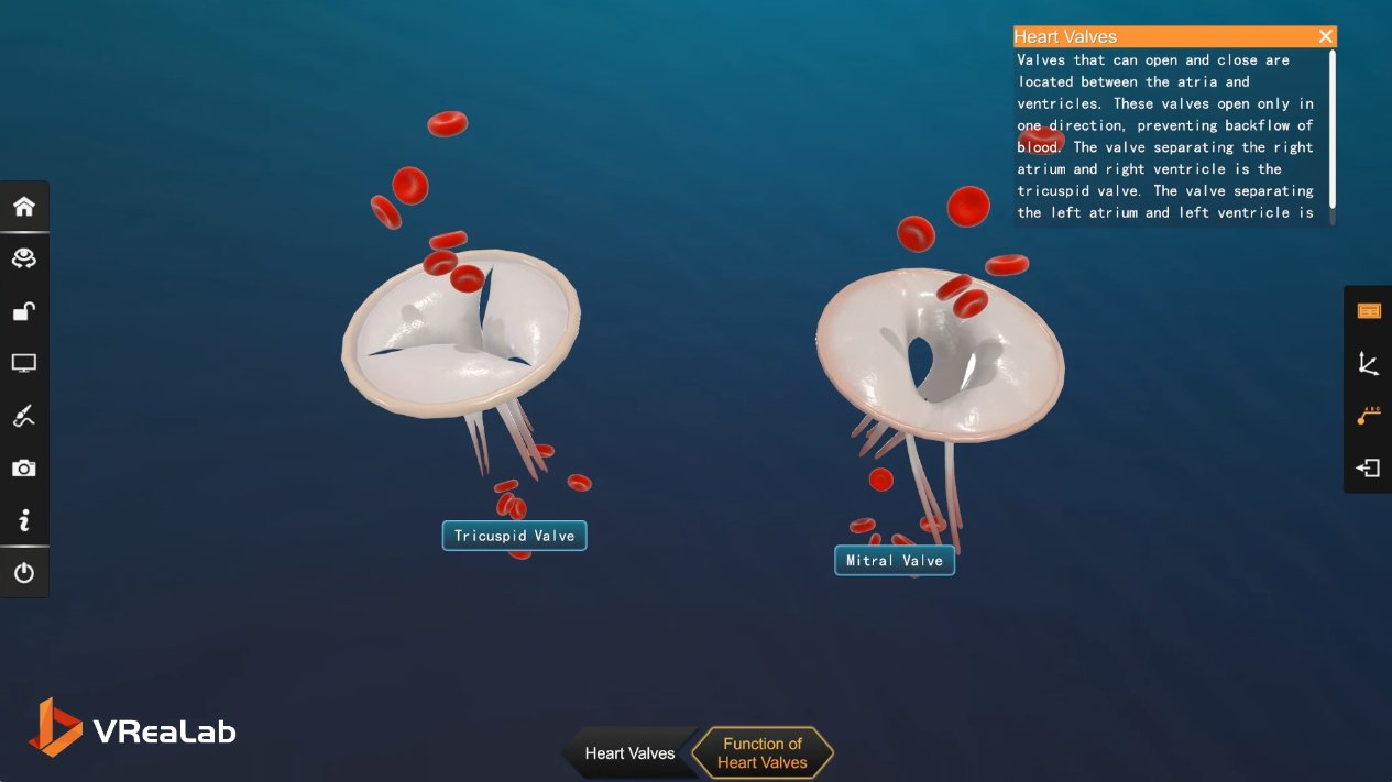
Figure 3. The tricuspid and mitral valves shown with red blood cells, demonstrating one-way flow and the prevention of backflow.
Following the Blood: Understanding the Heart at Work
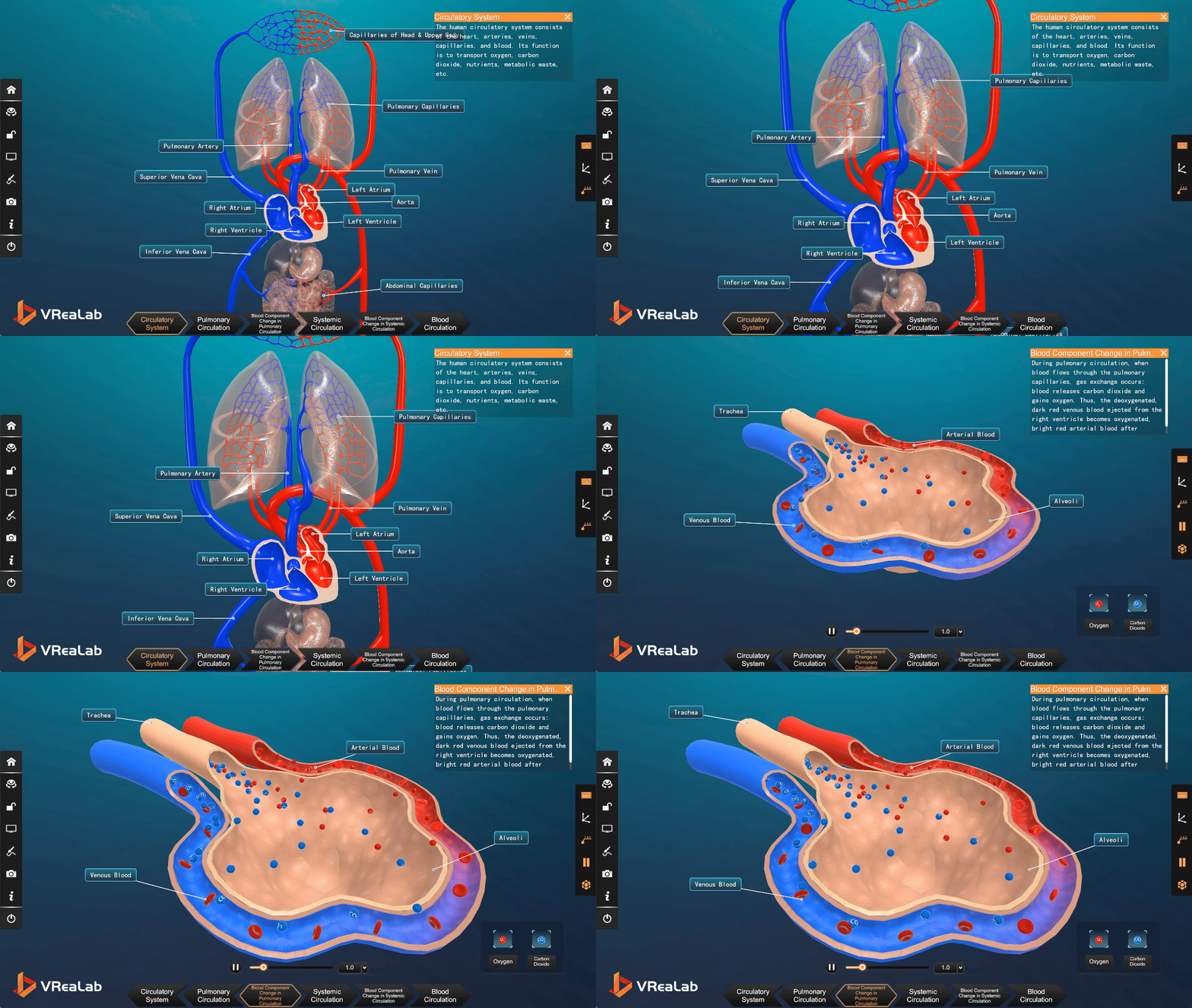
The value of the heart lies not only in its own beating but in its support of the entire circulatory system. In the VR learning process, learners can follow the perspective of the blood itself: pumped out of the heart into the aorta, carrying oxygen and nutrients to tissues throughout the body, then returning through the venous system to complete a full cycle. This follow-along experience turns the abstract pathway of blood circulation into something intuitive and concrete. Students can see the pulmonary circulation and the systemic circulation as one connected journey within the complete Human Body Systems 3D Model, and they come to understand why the heart is called the power pump of the human body.
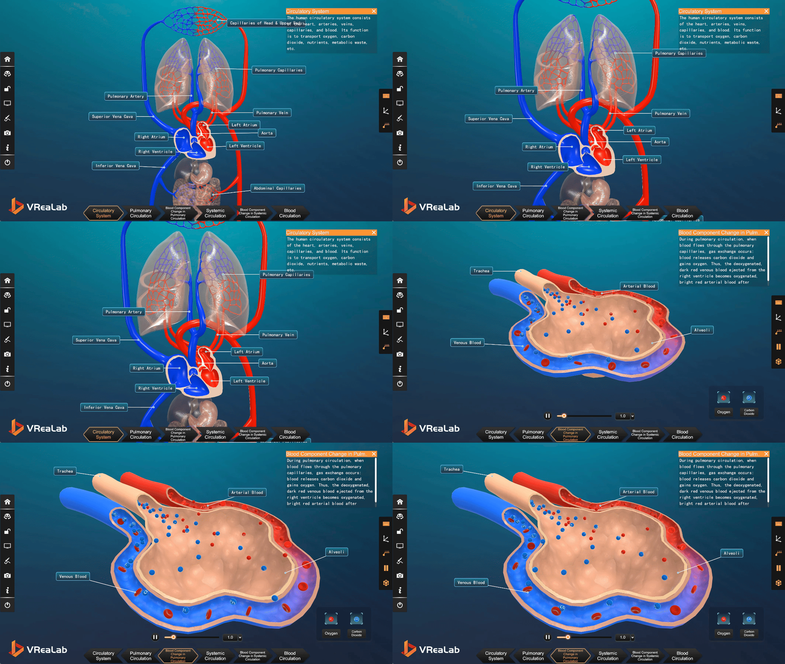
Figure 4. The full blood circulation system: pulmonary and systemic circuits connected through the four chambers of the heart.
A Safe and Repeatable Virtual Experiment
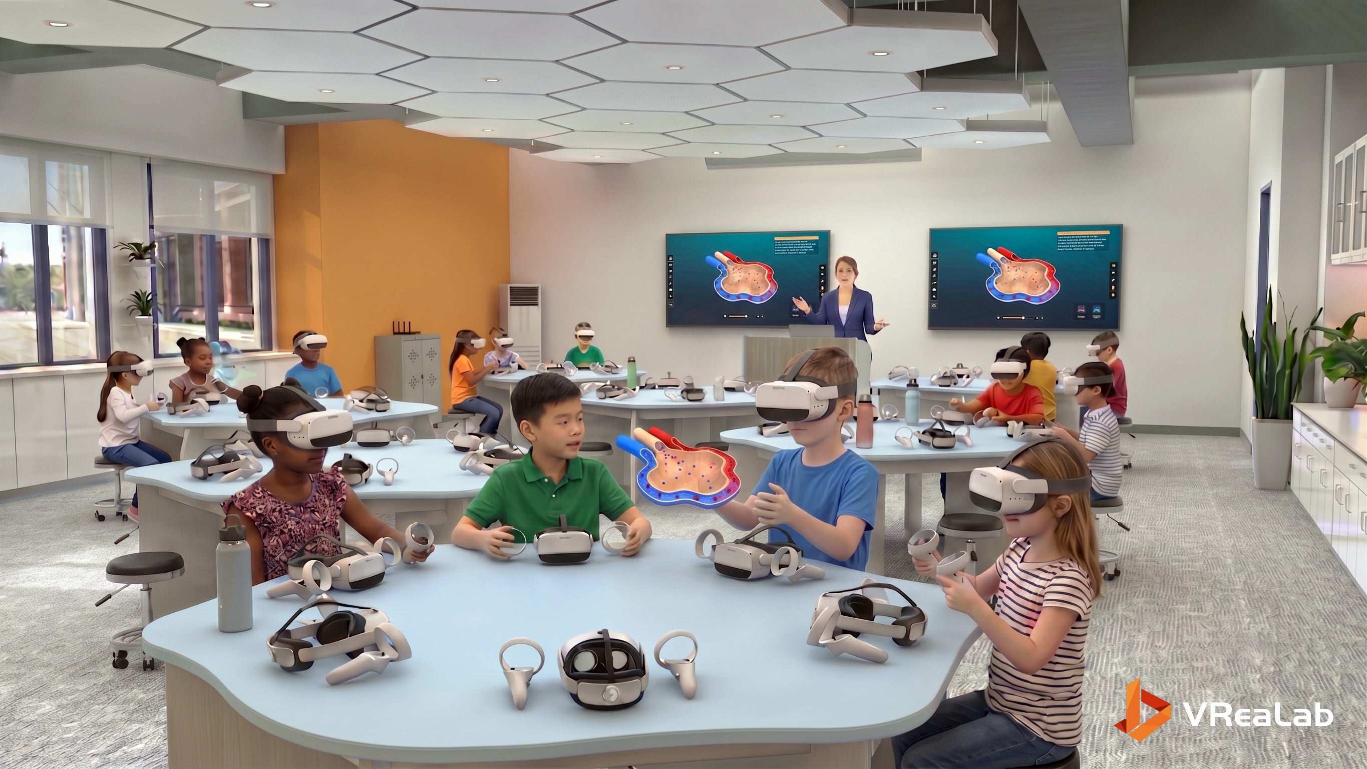
In real classrooms, human dissection and physiological experiments are limited by conditions, safety, and ethics, and can rarely be carried out fully or repeatedly. The virtual simulation laboratory removes these barriers. Students can observe the same structure as many times as needed, adjust the viewing angle to discover new details, and study the internal systems of the human body in depth without any ethical risk. This safe, repeatable, magnifiable way of learning is a powerful supplement to physiology teaching in primary and secondary schools. It also changes the role of the teacher: instead of drawing the same diagram on the board year after year, the teacher guides exploration, poses questions, and lets students verify their own predictions inside the scene. When the heart can be opened, entered, and replayed at will, understanding finally replaces memorization. Supported by a rigorous 3d anatomy of the human body and sustained biology interactive learning, every student carries away a living picture of the system that keeps them alive.
Source: Word document uploaded by user